Wednesday, February 28, 2007

Institutional Review Boards and Open Access Publishing

The New York Times has an article about whether the rules for Human Subjects protection, enforced by Institutional Review Boards, have in fact gone overboard (in terms of onerous regulations that protect no one):
As Ethics Panels Expand Grip, No Field Is Off Limits

By PATRICIA COHEN

Ever since the gross mistreatment of poor black men in the Tuskegee Syphilis Study came to light three decades ago, the federal government has required ethics panels to protect people from being used as human lab rats in biomedical studies. Yet now, faculty and graduate students across the country increasingly complain that these panels have spun out of control, curtailing academic freedom and interfering with research in history, English and other subjects that poses virtually no danger to anyone.

The panels, known as Institutional Review Boards, are required at all institutions that receive research money from any one of 17 federal agencies and are charged with signing off in advance on almost all studies that involve a living person, whether a former president of the United States or your own grandmother. This results, critics say, in unnecessary and sometimes absurd demands.

Among the incidents cited in recent report by the American Association of University Professors are a review board asking a linguist studying a preliterate tribe to "have the subjects read and sign a consent form," and a board forbidding a white student studying ethnicity to interview African-American Ph.D. students "because it might be traumatic for them."
Of course, those who conduct research with human participants, whether in the biomedical sciences, psychology, or sociology, should adhere to guidelines that protect the rights of the individuals involved, as outlined in The Nuremberg Code, the Declaration of Helsinki, and the Belmont Report. However, as nearly everyone in research can attest, having your project held up for months because of changes like this...

(1) Results of the study to date – line f was changed from “total men and women minorities enrolled” to “minorities” as requested.

...is counterproductive!

[NOTE: the example above was taken from an actual cover letter for a revised protocol.]

This brings me to the next topic (which is related, you'll see): Open Access Publishing, illustrated by such journals as the Public Library of Science family. The concept of making the results of publically funded research freely available to all is a worthy one. The idea of charging a taxpayer $39.00 to read an article important to her health such as this:
Ozanne EM, Annis C, Adduci K, Showstack J, Esserman L. (2007). Pilot trial of a computerized decision aid for breast cancer prevention. Breast J. 13: 147-54

This study sought to evaluate a shared decision-making aid for breast cancer prevention care designed to help women make appropriate prevention decisions by presenting information about risk in context. The decision aid was implemented in a high-risk breast cancer prevention program and pilot-tested in a randomized clinical trial comparing standard consultations to use of the decision aid.
is an outlandish affront perpetrated by Blackwell Synergy, Elsevier, and other publishers (see press coverage).

One of the down sides of Open Access Publishing is the steep price that investigators must pay to publish their research. For instance, publication charges at PLoS Medicine (and others) are $2500! However,
We offer a complete or partial fee waiver for authors who do not have funds to cover publication fees. Editors and reviewers have no access to payment information, and hence inability to pay will not influence the decision to publish a paper.
But who (at a major research university) wants to admit they can't afford to pay?

Another problem is the journal policy of wanting to obtain signed patient consent forms!! For example, this from PLoS Clinical Trials:
Protocol Submission

If authors have published their trial protocol in a peer-reviewed journal, then they should simply cite it in their submission to PLoS Clinical Trials. If the original protocol has not been published separately, it should be provided on submission, ideally uploaded through the electronic manuscript submission system, as a supporting file. The protocol need not include case record forms, patient information leaflets, or consent forms, although we (and reviewers) reserve the right to see patient consent forms in particular circumstances. Authors should also provide any subsequent amendments from the original protocol.
This stipulation is in express violation of the consent forms themselves! The forms typically have a lengthy confidentiality clause, and PLoS's request of viewing signed consent forms, which contain protected information (i.e., the participant's name), is not allowed! So presumably, here they mean just a blank copy of the form. However,
Our policy conforms to the Uniform Requirements, which states:

"Patients have a right to privacy that should not be infringed without informed consent. Identifying information should not be published in written descriptions, photographs, and pedigrees unless the information is essential for scientific purposes and the patient (or parent or guardian) gives written informed consent for publication. Informed consent for this purpose requires that the patient be shown the manuscript to be published. Complete anonymity is difficult to achieve, and informed consent for publication should be obtained if there is any doubt. If data are changed to protect anonymity, authors should provide assurance that alterations of the data do not distort scientific meaning. When informed consent has been obtained it should be indicated in the published article."

In the unusual situation that a manuscript reports individual patient information that might potentially identify them, informed consent of the patient(s) will be required. Please use this form to obtain the patient's permission, and FAX to the PLoS UK office. PLoS Clinical Trials' open access license means that the images and text we publish online become available for any lawful purpose. This information should be conveyed when obtaining consent for publication from patients.
Now this stipulation does require submission of signed consent forms. Of course, you don't want to go around publishing identifying information from your participants, but the way PLoS defines "complete anonymity" is rather liberal (e.g., "Patient X is a 53 year old male..." would be too specific).

Any ideas or opinions on these matters?

Monday, February 26, 2007

Deep Brain Stimulation For Phantom Limb Pain

A clinical study of deep brain stimulation for the treatment of phantom limb pain (Bittar et al., 2005) reviews not only the treatments and mechanisms of phantom limb pain, but the historical aspects as well:
The 16th century French military surgeon Ambroise Pare was the first to medically document the phenomena of phantom limb sensation and phantom limb pain in 1551:

"For the patients, long after the amputation is made, say they still feel pain in the amputated part. Of this they complain strongly, a thing worthy and almost incredible to people who have not experienced this"

In 1830, the neurologist Charles Bell published a description of the condition in The Nervous System of the Human Body. The term ‘phantom’, however, is often credited to the American military surgeon Silas Weir Mitchell, who in 1871 gave the first modern description of a post-surgical ‘ghost’ occurring in an amputee:

"There is something almost tragical, something ghastly, in the notion of these thousands of spirit limbs haunting as many good soldiers, and every now and then tormenting them... when... the keen sense of the limb’s presence betrays the man into some effort, the failure of which of a sudden reminds him of his loss"
Bittar RG, Otero S, Carter H, Aziz TZ. (2005). Deep brain stimulation for phantom limb pain. J Clin Neurosci. 12: 399-404.

Phantom limb pain is an often severe and debilitating phenomenon that has been reported in up to 85% of amputees. Its pathophysiology is poorly understood. Peripheral and spinal mechanisms are thought to play a role in pain modulation in affected individuals; however central mechanisms are also likely to be of importance. The neuromatrix theory postulates a genetically determined representation of body image, which is modified by sensory input to create a neurosignature. Persistence of the neurosignature may be responsible for painless phantom limb sensations, whereas phantom limb pain may be due to abnormal reorganisation within the neuromatrix. This study assessed the clinical outcome of deep brain stimulation of the periventricular grey matter and somatosensory thalamus for the relief of chronic neuropathic pain associated with phantom limb in three patients. These patients were assessed preoperatively and at 3 month intervals postoperatively. Self-rated visual analogue scale pain scores assessed pain intensity, and the McGill Pain Questionnaire assessed the quality of the pain. Quality of life was assessed using the EUROQOL EQ-5D scale. Periventricular gray stimulation alone was optimal in two patients, whilst a combination of periventricular gray and thalamic stimulation produced the greatest degree of relief in one patient. At follow-up (mean 13.3 months) the intensity of pain was reduced by 62% (range 55-70%). In all three patients, the burning component of the pain was completely alleviated. Opiate intake was reduced in the two patients requiring morphine sulphate pre-operatively. Quality of life measures indicated a statistically significant improvement. This data supports the role for deep brain stimulation in patients with phantom limb pain. The medical literature relating to the epidemiology, pathogenesis, and treatment of this clinical entity is reviewed in detail.

Phantom Limb


The Shins

For a more accurate musical take on Phantom Limbs, see Men in a War (and listen to the song by Suzanne Vega).

Tuesday, February 20, 2007

alternative time consciousness platform

Here's a really alternative conception of time...

Reproduced from THE ORDER OF TIME.

FILOGNOSY

Statement of Purpose


The purpose of this site is to give clear and unequivocal instruction on the filognosy as proposed by
The Order of Time. The Order of Time proposes for the science, politics, spirituality, personal interest, art and surfing of an alternative of time-consciousness. For that purpose it has developed e-books, designs, articles, definitions, timetables, a humanities directory for searching the net and even a game for searching on your identity for your life's mission. The complexity and diversity of The Order of Time might obscure the simplicity of its basic premises. To be clear about this simplicity this site offers straight questions and answers of a basic nature that will explain the concepts of consciousness, discipline, duality, liberation, order etc.

There's also a book of rules:
The Other Rules
How to be in love with the world (and her)
These rules cover not only Daily Life, but also Dating, Relations (rule y: Avoid free sex, intoxicants, financial speculation and the use of animal), Sport (rule j: Always shower), and most importantly, Making Love:
h) If you think you don't need: accept things going bad.
Sex is, once conditioned to it a need. It makes a balance of neurotransmitters in the brain, a balance of energy and foodintake in the body, a balance of associations in society and a certain type of duty to the Godhead of worship, the political leader, scientific paradigm or even a climate. Sometimes the fix of sexual behavior is called life, while others call it a kind of death as nothing seems to happen outside the line of sexual conditioning: one does not really develop other interests or intelligence. Also creativity can be seriously blocked attached to certain perversions finding no resistance or genius.

v) Care for love-play after the climax.
Just as important not to be egoistically after sexual gratification it is also important to share the success of achieving on a higher level of discharge. Many people simply use others at the 'way to the top'. But the other is not simply a staircase or a sex-object to be discarded after success. It is because of the support of the government, the spouse, the teachers, the religion, the culture of whatever that one could achieve. Gratitude is a very important value in the appreciation of as well the sexual orgasm as a societal success. ... Sexual orgasm and societal success go together also although it is somewhat of a paradox to be a cultural success and a intimate success at the same time. Usually success of sexual people is made by celibates working behind the screen. [MY EMPHASIS]

The copyrights of the materials at this site are settled thus that each may use and copy them freely, provided it is done without a profit-motive.

Time Is A Delusion


Time, if we can intuitively grasp such an identity, is a delusion: the difference and inseparability of one moment belonging to its apparent past from another belonging to its apparent present is sufficient to disintegrate it.

-- Jorge Luis Borges, A New Refutation of Time

Monday, February 19, 2007

Present Tense


Lately, The Neurocritic (and recent neuronews) has been focused on the past and the future. But what about the present? What happens to the brain when we are truly focused on the present moment? Overlapping brain areas activated while remembering the past/imagining the future include medial prefrontal cortex, posterior cingulate, and parahippocampal gyrus (Okuda et al., 2003; Addis et al., 2007; Szpunar et al., 2007). Are these regions (and others specific to past or future) deactivated when one resides in the present moment? And what does this state entail, phenomenologically speaking? Mindfulness is now a very popular topic of academic study, and numerous universities have centers for research on mindfulness and the application of mindfulness techniques in clinical practice.

The Centre For Mindfulness Research And Practice (University of Wales, Bangor)

Center for Mindfulness in Medicine, Health Care, and Society (University of Massachusetts Medical School)

Mindfulness Practice Center (University of Vermont)

UCSD Center for Mindfulness

etcetera...

What is mindfulness? According to Wikipedia, mindfulness is
the practice whereby a person is intentionally aware of his or her thoughts and actions in the present moment, non-judgmentally. Mindfulness is applied to both bodily actions and the mind's own thoughts and feelings.
One of the pioneers in this area is Richie Davidson, director of the Laboratory for Affective Neuroscience at the University of Wisconsin. Certainly, this topic can cover an entire blog, but in terms of neuroimaging studies, the literature on mindfulness-based meditation is sparse. However, two preliminary experiments were reported at conferences and summarized by Cahn and Polich (2006) in their comprehensive review, Meditation States and Traits: EEG, ERP, and Neuroimaging Studies. One of these fMRI studies (Baerentsen, 2001), and another one by Ritskes et al. (2003), employed 45 sec on/45 sec off bouts of meditation/rest (not entirely conducive to entering the appropriate state). At any rate, Baerentsen (2001) reported activations in the dorsolateral prefrontal cortex (PFC), anterior cingulate cortex (ACC), hippocampus, and right temporal lobe, and deactivations in the visual cortex. Ritskes et al. (2003) also reported increased activity in dorsolateral PFC (R>L) plus the basal ganglia. They saw decreased activity in the occipital lobe but also in the anterior cingulate (in contrast to Baerentsen, 2001). Hmm. Anyway. Commonalities include deactivation in the occipital cortex (which is activated in the past/future studies, presumably due to imagery) and activation in dorsolateral PFC.

At first glance, the latter finding seems curious because dorsolateral PFC is involved in attention, working memory, and other executive control-type functions. Although meditation is relaxing, it is
a state of concentrated attention on some object of thought or awareness.
This point was stressed by Lazar and colleagues (2000), who reported greater activity in dorsolateral PFC, parietal lobe, ACC, striatum, hippocampus/parahippocampus, etc. during the practice of Kundalini meditation, which involves a focus on breathing and silent repetition of phrases during inhalation and exhalation.

Another unpublished study compared two forms of meditation: Kundalini (mantra-based) or Vipassana (mindfulness-based). These were contrasted with control conditions of rest, random number generation, and paced breathing (Lazar et al., 2003). Similar (but nonoverlapping) regions in frontal and parietal cortices and some subcortical areas were more active during the two types of meditation than during the control conditions. A common area of activation was in the dorsal ACC. Overall, these findings suggest a focused state of attention during meditation, which engages an attentional network (mostly) unlike what is seen in studies of past and future time travel. Areas common to reflecting upon the past, present, and future include the hippocampus and parahippocampal gyrus.

References

Addis DR, Wong AT, Schacter DL. (2007). Remembering the past and imagining the future: Common and distinct neural substrates during event construction and elaboration. Neuropsychologia 45: 1378-1385.

Baerentsen KB. (2001). Onset of meditation explored with fMRI. Neuroimage 13: S297.

Cahn BR, Polich J (2006). Meditation States and Traits: EEG, ERP, and Neuroimaging Studies. Psych Bull. 132: 180-211.

Lazar SW, Rosman IS, Vangel M, Rao V, Dusek, H, Benson H, et al. (2003, November). Functional brain imaging of mindfulness and mantra-based meditation. Paper presented at the meeting of the Society for Neuroscience, New Orleans, LA.

Lazar SW, Bush G, Gollub RL, Fricchione GL, Khalsa G, Benson H. (2000). Functional brain mapping of the relaxation response and meditation. NeuroReport 11: 1581-1585.

Okuda J, Fujii T, Ohtake H, Tsukiura T, Tanji K, Suzuki K, et al. (2003). Thinking of the future and the past: The roles of the frontal pole and the medial temporal lobes. Neuroimage 19: 1369–1380.

Ritskes, R., Ritskes-Hoitinga, M., Stodkilde-Jorgensen, H., Baerentsen, K., & Hartman, T. (2003). MRI scanning during Zen meditation: The picture of enlightenment? Constructivism in the Human Sciences 8: 85-90.

Szpunar KK, Watson JM, McDermott KB. (2007). Neural substrates of envisioning the future. Proc. Natl. Acad. Sci. Published online before print January 3, 2007.


Sunday, February 18, 2007

past present future


We're here, there, not here, not there, swirling like specks of dust, claiming for ourselves the rights of the universe. Being important, being nothing, being caught in lives of our own making that we never wanted. Breaking out, trying again, wondering why the past comes with us, wondering how to talk about the past at all.

--Jeanette Winterson, Lighthousekeeping

Saturday, February 10, 2007

Fables Of The Reconstruction


A flurry of papers (well, OK, three) has been published recently on the relationship between how we remember the past and imagine the future. The third and final paper is another functional neuroimaging study (Addis et al., 2007).

Before we begin, let’s highlight a 2003 study by Okuda and colleagues. They were the first to compare the neural correlates of imagining past and future events. Common regions engaged by both forms of (re)construction included bilateral medial PFC, hippocampus, parahippocampal gyrus, and the left precuneus (Okuda et al., 2003). Looks fairly similar to the Bill Clinton study (Szpunar et al., 2007), except the latter lacked hippocampal activation and saw future > past for left precuneus.

So why is it big news now, when it’s not novel? That’s a question for sociologists to answer…


Anyway, Addis et al. did point out that the older study used PET and a blocked design where participants spoke freely about the past or the future during a 60 sec time interval (Okuda et al., 2003), meaning there was no temporal precision to link specific brain activations to specific events. So similar to Szpunar et al., the Addis experimental design consisted of giving cue words to prompt either remembered or imagined events. The control conditions, however, were quite different: semantic retrieval (think of two words related to the cue word and then use all three words in a sentence) and visual imagery (think of two objects related to the cue word, one larger and one smaller). In addition, they devised a rating scale for

the episodic specificity of events generated during scanning, and collected subjective ratings of the level of detail, emotionality, personal significance and field/observer perspective.

They also tried to separate the participants’ introspection about the past and future into a construction phase and an elaboration phase.

Fitting for such a complicated design, the results were complicated. First, there was a set of regions more active for event construction (past+future) than for control construction (semantic+imagery), shown in (a) below. Then there was another set of regions more active for event elaboration than for control elaboration, shown in (b) below. So these brain areas were hoppin’ to an equivalent extent for past and future.

Then the future and the past were compared directly. There seemed to be no regions that were more active for (supposedly) veridical memory than for imagination. Hmm. There were a bunch of areas with greater activity for prospective event construction (i.e., future) than for historical reconstruction (i.e., past), as seen in (a) below.

But what about memory retrieval??

In contrast to common past–future activity in the left hippocampus, the right hippocampus was differentially recruited by future event construction. This finding is notable, not only because others report right hippocampal activity to be common to both past and future events (Okuda et al., 2003) but also because it is surprising that future events engage a structure more than the very task it is thought to be crucial for: retrieval of past autobiographical events…

It does seem strange that no regions were more active for memory than for imagination. So memory doesn’t differ from fiction? At the very least, it didn’t result in greater brain activity than fiction, not in this particular study (an important point).

There was no evidence of any regions engaged uniquely by past events, not only in the PFC but across the entire brain. This outcome was unexpected in light of previous results (Okuda et al., 2003). Moreover, regions mediating retrieval processes (e.g., cue-specification, Fletcher et al., 1998) such right ventrolateral PFC (e.g., BA 47) should be engaged by a pure retrieval task (i.e., past events) more than a generation task (i.e., future events). More surprising was the finding that right BA47 showed more activity for future than past events, and that past events did not engage this region significantly more than control tasks.

I won’t try to summarize how the authors interpret the rest of their data, so you’ll have to read the abstract.

Addis DR, Wong AT, Schacter DL. (2007). Remembering the past and imagining the future: Common and distinct neural substrates during event construction and elaboration. Neuropsychologia 45: 1378-1385.

People can consciously re-experience past events and pre-experience possible future events. This fMRI study examined the neural regions mediating the construction and elaboration of past and future events. Participants were cued with a noun for 20 s and instructed to construct a past or future event within a specified time period (week, year, 5–20 years). Once participants had the event in mind, they made a button press and for the remainder of the 20 s elaborated on the event. Importantly, all events generated were episodic and did not differ on a number of phenomenological qualities (detail, emotionality, personal significance, field/observer perspective). Conjunction analyses indicated the left hippocampus was commonly engaged by past and future event construction, along with posterior visuospatial regions, but considerable neural differentiation was also observed during the construction phase. Future events recruited regions involved in prospective thinking and generation processes, specifically right frontopolar cortex and left ventrolateral prefrontal cortex, respectively. Furthermore, future event construction uniquely engaged the right hippocampus, possibly as a response to the novelty of these events. In contrast to the construction phase, elaboration was characterized by remarkable overlap in regions comprising the autobiographical memory retrieval network, attributable to the common processes engaged during elaboration, including self-referential processing, contextual and episodic imagery. This striking neural overlap is consistent with findings that amnesic patients exhibit deficits in both past and future thinking, and confirms that the episodic system contributes importantly to imagining the future.

Reference

Okuda J, Fujii T, Ohtake H, Tsukiura T, Tanji K, Suzuki K, et al. (2003). Thinking of the future and the past: The roles of the frontal pole and the medial temporal lobes. Neuroimage 19: 1369–1380.

Read Part 1 and Part 2.


The Reconstruction of Fables
Appendix A. Example of a specific future event generated by a pilot participant.

Future event (in 5 years; cue=dress)

My sister will be finishing . . . her undergraduate education, I imagine some neat place, Ivy league private school . . . it would be a very nice spring day and my mom and my dad will be there, my dad with the camcorder as usual, and my mom with the camera as usual. My sister will be in the crowd and they’d be calling everyone’s name . . . I can see her having a different hair style by then, maybe instead of straight, very curly with lots of volume. She would be wearing contacts by then and heels of course. And I can see myself sitting in some kind of sundress, like yellow, and under some trees . . . the reception either before or after and it would be really nice summer food, like salads and fruits, and maybe some sweets, and cold drinks that are chilled but have no ice. And my sister would be sitting off with her friends, you know, talking with them about graduating, and they’d probably get emotional.

Wednesday, February 7, 2007

i miss you but i haven't met you yet

i miss you
but i haven't met you yet
so special
but it hasn't happened yet
you are gorgeous
but i haven't met you yet
i remember
but it hasn't happened yet


Remembering The Past, Envisioning The Future

As Björk sings about her temporal conundrum, let's continue our time tunnel from the other day. Two recent neuroimaging papers have related the past to the future, as far as the brain is concerned. In the first study (Szpunar et al., 2007), participants were presented with a bunch of cue words (e.g., birthday, getting lost, etc) in the scanner and were told to orient their attention to the cue event. Then they had to either think about the event in relation to their future (SF), in relation to their past (SR), or in relation to a familiar individual (CI). The latter was a snappy and fun control condition: imagine Bill Clinton in that situation! OK, so comparisons were made between self-remember (SR), self-future (SF), and Clinton-imagine (CI). A set of regions (A-D), illustrated below in the left side of the figure, were more active for the future than the past. Another set of regions, illustrated on the right, had statistically equivalent activation when participants remembered the past and imagined the future.


from Szpunar et al. (2007)

Future > Past

Future = Past

(A) L Middle Frontal

(E) Medial Frontal

(B) L Middle Frontal

(F) Parahippocampal Gyrus

(C) L Precuneus

(G) Posterior Cingulate

(D) R Cerebellum

(H) L Superior Occipital


The regions that were more active in imaging one's own future (not Bill's or Hillary's) included left lateral premotor cortex, our friend the precuneus in the left hemisphere, and the right cerebellum. [Note that left frontal cortex and right cerebellum are interconnected (Leiner et al., 1989; Niimura et al., 1999; Jansen et al., 2005]. The authors speculate that activation in a circuit that represents imagined motor movements is greater for novel movements that haven't occurred yet than for remembered motor sequences... hmmm.

Then there was a set of regions equally active when placing oneself in the past and in the future. These included elements of the dreaded default network (medial prefrontal, posterior cingulate). In fact, for the medial prefrontal ROI in panel E above, there was "less deactivation" (actually, the hemodynamic response looks flat and at zero) for self-related conditions than for the Clinton condition. More interesting are the other 3 regions: the parahippocampal gyrus (part of the medial temporal lobe memory system and home to the parahippocampal place area), posterior cingulate, and occipital cortex. The authors link these areas to autobiographical memories and spatial navigation. And perhaps, to Endel Tulving's conception of autonoetic consciousness, the ability to mentally represent and become aware of subjective experiences in the past, present, and future.


Karl K. Szpunar, Jason M. Watson, and Kathleen B. McDermott. Neural substrates of envisioning the future. Proc. Natl. Acad. Sci. Published online before print January 3, 2007. OPEN ACCESS ARTICLE.

The ability to envision specific future episodes is a ubiquitous mental phenomenon that has seldom been discussed in the neuroscience literature. In this study, subjects underwent functional MRI while using event cues (e.g., Birthday) as a guide to vividly envision a personal future event, remember a personal memory, or imagine an event involving a familiar individual. Two basic patterns of data emerged. One set of regions (e.g., within left lateral premotor cortex; left precuneus; right posterior cerebellum) was more active while envisioning the future than while recollecting the past (and more active in both of these conditions than in the task involving imagining another person). These regions appear similar to those emerging from the literature on imagined (simulated) bodily movements. A second set of regions (e.g., bilateral posterior cingulate; bilateral parahippocampal gyrus; left occipital cortex) demonstrated indistinguishable activity during the future and past tasks (but greater activity in both tasks than the imagery control task); similar regions have been shown to be important for remembering previously encountered visual-spatial contexts. Hence, differences between the future and past tasks are attributed to differences in the demands placed on regions that underlie motor imagery of bodily movements, and similarities in activity for these two tasks are attributed to the reactivation of previously experienced visual-spatial contexts. That is, subjects appear to place their future scenarios in well known visual-spatial contexts. Our results offer insight into the fundamental and little-studied capacity of vivid mental projection of oneself in the future.
From Behavioral Neuroscience 103: 998-1008, October 1989.


Parallel cerebello-frontal connections in the human brain. (Different parts of the cerebellum are linked to different areas of the frontal lobe. Because these connections enable the frontal areas to send signals to the cerebellum and receive signals from it, the cerebellum can serve as an information-processing adjunct to all of these frontal areas. Information can be processed repeatedly in such cerebro-cerebellar loops during the waking hours of life.)

This article generated a lot of press coverage, and some of it was actually fairly decent.
Scan shows how brains plot future

The Washington University team say that specific areas of the brain are active when thinking about upcoming events.

. . .

The researchers placed 21 volunteers into the MRI machine, then contrasted the scan results when they were asked to imagine vividly future events and recollect past memories.

The resulting images showed clear differences between a birthday already experienced, and a birthday yet to come.

In particular, when looking ahead, three particular areas of the brain were activated - the left lateral premotor cortex, the left precuneus and the right posterior cerebellum.

These brain areas are already known to be involved in the imagining of body movements, suggesting that when the human brain is thinking about the future, it does so in terms of distinct movements and actions that will happen at that point.
Brain Uses Past to Peer Into Future

References

Jansen A, Floel A, Van Randenborgh J, Konrad C, Rotte M, Forster AF, Deppe M, Knecht S. (2005). Crossed cerebro-cerebellar language dominance. Hum Brain Mapp. 24: 165-72.

Leiner HC, Leiner AL, Dow RS. (1989). Reappraising the cerebellum: what does the hindbrain contribute to the forebrain? Behav Neurosci.103: 998-1008.

Niimura K, Chugani DC, Muzik O, Chugani HT. (1999). Cerebellar reorganization following cortical injury in humans: effects of lesion size and age. Neurology 52: 792-7.

Memory Expert's Memory About False Memory Is False

Yes, this one isn't "Just Science," and it's over 3 months old, but it fits in with the recent memory theme here at The Neurocritic.
In the Libby Case, A Grilling to Remember
The Washington Post. Washington, D.C.:Oct 27, 2006. p. A.21
. . .

Fitzgerald's target in the witness box was Elizabeth F. Loftus, a professor of criminology and psychology at the University of California at Irvine. For more than an hour of the pretrial hearing, Loftus calmly explained to Judge Reggie B. Walton her three decades of expertise in human memory and witness testimony. Loftus asserted that, after copious scientific research, she has found that many potential jurors do not understand the limits of memory and that Libby should be allowed to call an expert to make that clear to them.

But when Fitzgerald got his chance to cross-examine Loftus about her findings, he had her stuttering to explain her own writings and backpedaling from her earlier assertions. Citing several of her publications, footnotes and the work of her peers, Fitzgerald got Loftus to acknowledge that the methodology she had used at times in her long academic career was not that scientific, that her conclusions about memory were conflicting, and that she had exaggerated a figure and a statement from her survey of D.C. jurors that favored the defense.

Her defense-paid visit to the federal court was crucial because Libby is relying on the "memory defense" against Fitzgerald's charges that he obstructed justice and lied to investigators about his role in the leaking of a CIA operative's identity to the media. Libby's attorneys argue that he did not lie -- that he was just really busy with national security matters and forgot some of his conversations.

When Fitzgerald found a line in one of her books that raised doubts about research she had cited on the stand as proof that Libby needs an expert to educate jurors, Loftus said, "I don't know how I let that line slip by."

"I'd need to see that again," Loftus said when Fitzgerald cited a line in her book that overstated her research by saying that "most jurors" consider memory to be equivalent to playing a videotape. Her research, however, found that to be true for traumatic events, and even then, only 46 percent of potential jurors thought memory could be similar to a videotape.

There were several moments when Loftus was completely caught off guard by Fitzgerald, creating some very awkward silences in the courtroom.

One of those moments came when Loftus insisted that she had never met Fitzgerald. He then reminded her that he had cross-examined her before, when she was an expert defense witness and he was a prosecutor in the U.S. attorney's office in New York.

Monday, February 5, 2007

Mental Time Travel


"The Time Tunnel" (1966)

Endel Tulving, that Great Canadian Psychology Researcher and memory theorist, has long held that episodic memory, or the vivid recollection of events and their associated spatiotemporal contextual details, affords humans the unique ability to engage in mental time travel:
What makes mental time travel possible?

Psychologist Endel Tulving offered a theory on our uniquely human ability to act today based on our past and future.

BY BRIDGET MURRAY

Remembering, to most of us, means recalling a past occurrence. But to Endel Tulving, PhD, the mechanisms of memory evoke the future as well. The reason? Memory allows us to mentally travel backward in time as well as into the future, explained Tulving, a University of Toronto professor emeritus and visiting professor in cognitive neuroscience at Washington University, in a presidential invited address at APA's 2003 Annual Convention in Toronto.

Tulving's theory stems from extensive memory research he's conducted since the 1950s at Toronto, Yale University and the Toronto-based Rotman Research Institute--and, he said, others' research supports it too. He proposed an official term for, and definition of, what makes such mental time travel possible:

Chronesthesia--A hypothetical brain/mind ability or capacity, acquired by humans through evolution, that allows them to be constantly aware of the past and the future.
As a side note, the view that chronesthesia (Kim, 2007) or autonoetic consciousness (Wheeler et al., 1995), or ecphory (Kapur et al., 1995; Steinvorth et al., 2006) are distinctly human abilities has not gone without debate (Clayton et al., 2003; Dere et al., 2006; Emery et al., 2004; Suddendorf et al., 1997; Zentall, 2006).

So what's new and exciting? Here's a question: must we be able remember the past to envision the future? The answer is yes, according to a recent study conducted in amnesic patients with damage to the hippocampus (Hassabis et al., 2007). As reported in Science's News of the Week:
A Surprising Connection Between Memory and Imagination

Greg Miller

According to a new study, people with amnesia caused by damage to the hippocampus, a brain region intimately tied to memory, have difficulty envisioning commonplace scenarios they might reasonably expect to encounter in the future. The findings challenge long-held views about the function of the hippocampus and the nature of memory.
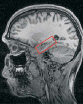
The Janus center? The hippocampus (red box) may be as important for imagining the future as it is for remembering the past. CREDIT: ELEANOR MAGUIRE

More press coverage:

Amnesiacs May Be Cut Off From Past and Future Alike

. . . The results suggest that to the brain, remembered experience and imagined experience are reflections from the same mirror, rich inner worlds animated by almost identical neural networks.


Hmm, aren't these findings exaggerated just a little bit, Mr. Carey? Fortunately, one of the authors puts the results into a better context:
"We think that what the hippocampus provides is a scaffold for experience and imagination, and that scaffold is spatial," Maguire said. The brain's record of physical space, she said, appears to be necessary to infuse a scene with rich personal dimension.
Five people with amnesia participated in the study. These individuals had contracted limbic encephalitis or meningitis that resulted in brain damage that appeared to extend to regions beyond the hippocampus proper. Memory loss was not only anterograde (an inability to encode new information) but also substantially retrograde (loss of remote autobiographical memories), extending from 10 years premorbidly to the entire lifetime for one person. These folks are not your classic cases of hippocampal amnesia (e.g., see Bayley et al., 2006), because their retrograde memory impairments are much more severe. This blows up yet another controversy swirling around the importance of the hippocampus in the retrieval of remote memories (important: Nadel & Moscovitch, 1997; not: Bayley et al., 2003).

The amnesic and control participants were given cue sentences (e.g., "Imagine you're lying on a white sandy beach in a beautiful tropical bay") and asked to vividly imagine such a scene, describing it using as much detail as possible. They were told not to recount an actual memory, but to create something new. [NOTE: how do you verify that non-amnesic subjects avoided describing actual memories of such a scene?] At any rate, the amnesic patients did, indeed, perform more poorly than controls on this task.
Hassabis D, Kumaran D, Vann SD, Maguire EA. (2007). Patients with hippocampal amnesia cannot imagine new experiences. Proc Natl Acad Sci 104: 1726-31.

Amnesic patients have a well established deficit in remembering their past experiences. Surprisingly, however, the question as to whether such patients can imagine new experiences has not been formally addressed to our knowledge. We tested whether a group of amnesic patients with primary damage to the hippocampus bilaterally could construct new imagined experiences in response to short verbal cues that outlined a range of simple commonplace scenarios. Our results revealed that patients were markedly impaired relative to matched control subjects at imagining new experiences. Moreover, we identified a possible source for this deficit. The patients' imagined experiences lacked spatial coherence, consisting instead of fragmented images in the absence of a holistic representation of the environmental setting. The hippocampus, therefore, may make a critical contribution to the creation of new experiences by providing the spatial context into which the disparate elements of an experience can be bound. Given how closely imagined experiences match episodic memories, the absence of this function mediated by the hippocampus, may also fundamentally affect the ability to vividly re-experience the past.

Back in 1978, John O'Keefe and Lynn Nadel published a book called The Hippocampus as a Cognitive Map. [now freely available!] In it, they built a theory of hippocampal function around the existence of place cells (O'Keefe & Dostrovsky, 1971) and theta activity (Vanderwolf, 1969). [We'll leave theta aside for now.] Place cells are hippocampal neurons that show increased firing rates whenever an animal is in a specific location in the environment - the cell's "place field."

Thus, the current results of Hassabis et al. emphasize the importance of spatial representations and implicate a different area of the brain for mental time travel than the one initially suggested by Tulving and colleagues in the 90's (e.g., Wheeler et al., 1997) -- namely, right hemisphere prefrontal cortex. No matter, Professor Tulving contributed the article for publication as a member of the National Academy of Science. At any rate, it seems clear that it takes a whole village of brain regions to envision the future and remember the past. And what of chronesthesia?
As for hard scientific evidence of chronesthesia's existence, there's "zero, none, very little--it's just an idea," said Tulving. But, he said, emerging imaging research promises to help shed light on its brain mechanisms and has already suggested that higher-order thinking regions, such as the prefrontal cortex, are involved.

He admonished, however, that no function--chronesthesia or any other--"holds a particular seat in the brain; it's all over the place."
You can also find some interesting commentary about the role of hippocampus in Imagination and memory over at Gene Expression.

References

Bayley PJ, Hopkins RO, Squire LR. (2003). Successful recollection of remote autobiographical memories by amnesic patients with medial temporal lobe lesions. Neuron 38: 135-44.

Bayley PJ, Hopkins RO, Squire LR. (2006). The fate of old memories after medial temporal lobe damage. J Neurosci. 26:13311-7.

Clayton NS, Bussey TJ, Dickinson A. (2003). Can animals recall the past and plan for the future? Nat Rev Neurosci 4: 685-91.

Dere E, Kart-Teke E, Huston JP, De Souza Silva MA. (2006). The case for episodic memory in animals. Neurosci Biobehav Rev 30:1206-24.

Emery NJ, Clayton NS. (2004). The mentality of crows: convergent evolution of intelligence in corvids and apes. Science 306: 1903-7.

Kapur S, Craik FI, Jones C, Brown GM, Houle S, Tulving E. (1995). Functional role of the prefrontal cortex in retrieval of memories: a PET study. Neuroreport 14:1880-4.

Kim, A (2007). Which brain regions enable us to remember our past and anticipate our future? Sci Consciousness Rev.

Nadel L, Moscovitch M. (1997). Memory consolidation, retrograde amnesia and the hippocampal complex. Curr Opin Neurobiol. 7: 217-27.

O'Keefe J, Dostrovsky J. (1971). The hippocampus as a spatial map. Preliminary evidence from unit activity in the freely-moving rat. Brain Res. 34:171-5.

O'Keefe J, Nadel L. (1978). The Hippocampus as a Cognitive Map. Oxford University Press.

Steinvorth S, Corkin S, Halgren E. (2006). Ecphory of autobiographical memories: an fMRI study of recent and remote memory retrieval. Neuroimage 30: 285-98.

Suddendorf T, Corballis MC. (1997). Mental time travel and the evolution of the human mind. Genet Soc Gen Psychol Monogr 123: 133-67.

Wheeler MA, Stuss DT, Tulving E. (1997). Toward a theory of episodic memory: the frontal lobes and autonoetic consciousness. Psychol Bull 121:331-54.

Zentall TR. (2006). Mental time travel in animals: a challenging question. Behav Processes 72: 173-83.